.svg)
Tell Us a Little More About Yourself!
Scrotal Ultrasound for Male Fertility and Urologic Evaluation
Scrotal ultrasound is one of the most valuable diagnostic tools in male urology and fertility care. It provides high-resolution, real-time imaging of the testicles and surrounding structures, allowing clinicians to identify conditions that cannot be diagnosed through physical examination alone.
At The Y Factor, scrotal ultrasound is used as a targeted diagnostic study, not as a routine or reflexive test. When ordered appropriately, it plays a critical role in understanding fertility challenges, evaluating pain or swelling, and identifying anatomic or vascular contributors that directly affect treatment decisions.

What Is a Scrotal Ultrasound?
A scrotal ultrasound is a non-invasive imaging study that uses sound waves to visualize the testicles, epididymis, spermatic cords, and surrounding tissues. Doppler imaging is often included to assess blood flow.
Unlike many tests, scrotal ultrasound provides objective structural and vascular information that cannot be inferred from lab tests alone and plays an important role in a diagnostic evaluation.

Why Scrotal Ultrasound Matters in Men’s Health
Many male reproductive and urologic conditions are structural or vascular in nature. These include:
- varicocele
- testicular asymmetry
- scrotal masses or cysts
- blood flow abnormalities
- post-inflammatory or post-injury changes
Without imaging, these contributors may go undetected, leading to incomplete diagnostic evaluation or inappropriate treatment. Scrotal ultrasound adds clarity and confidence to clinical decision-making.
What a Scrotal Ultrasound Can Evaluate
Testicular Anatomy and Structure
Ultrasound allows detailed evaluation of:
- testicular size and symmetry
- tissue consistency
- evidence of scarring or atrophy
Changes in testicular structure may reflect impaired sperm production, prior injury, or chronic conditions affecting fertility potential.
Blood Flow and Vascular Health
Doppler ultrasound evaluates blood flow to and from the testicles. Adequate blood flow is essential for:Vascular abnormalities may affect both fertility and testicular health.
- testicular function
- temperature regulation
- hormone production
Varicocele Detection
Varicoceles—dilated veins within the scrotum—are one of the most common reversible causes of male infertility. While large varicoceles may be detected on physical exam, smaller or subclinical varicoceles often require ultrasound for diagnosis.Ultrasound helps assess:
- presence and size of varicocele
- impact on testicular size
- laterality (left vs bilateral)
This information is essential for determining whether intervention may be beneficial.
Scrotal Masses and Fluid Collections
Ultrasound can distinguish between:
- solid vs cystic masses
- benign fluid collections
- inflammatory changes
This differentiation is critical for both reassurance and appropriate referral when needed.

Who Should Consider a Scrotal Ultrasound
Scrotal ultrasound may be recommended for men with:
- abnormal semen analysis results
- unexplained infertility
- testicular pain or swelling
- scrotal asymmetry
- suspected varicocele
- history of testicular injury or surgery
Imaging is particularly valuable when physical exam findings are subtle or unclear.
.webp)
Symptoms and Findings That Prompt Imaging
Common reasons for scrotal ultrasound include:
- persistent or recurrent scrotal pain
- sensation of heaviness or fullness
- fertility concerns without clear lab explanation
- visible or palpable scrotal changes
- follow-up of known conditions
Symptoms may be mild, but imaging can reveal meaningful contributors.
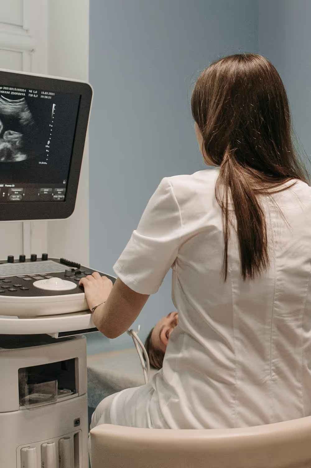
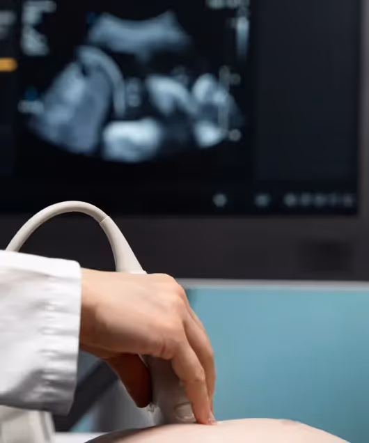
How a Scrotal Ultrasound Is Performed
The Ultrasound Procedure
The procedure is performed in-office or at an imaging facility and typically takes 15–30 minutes. A handheld transducer is used to capture images while the patient lies comfortably.
No needles, radiation, or sedation are involved.
Safety and Comfort Considerations
Scrotal ultrasound is:
- non-invasive
- painless
- safe
- repeatable
Most men tolerate the procedure well, and results are available quickly.
.avif)
Interpreting Scrotal Ultrasound Findings
Ultrasound findings must be interpreted in clinical context. Not all abnormalities require intervention. Interpretation considers:
- fertility goals
- symptom severity
- correlation with semen analysis
- progression over time
This prevents unnecessary treatment while identifying meaningful issues.
Scrotal Ultrasound and Male Fertility
For men facing fertility challenges, scrotal ultrasound helps identify:
- varicocele-related impairment
- testicular size discrepancies
- structural contributors to abnormal sperm parameters
Imaging results may guide decisions around observation, optimization, or referral for advanced reproductive planning.
Scrotal Ultrasound in Pelvic and Scrotal Pain
Pain in the scrotum may arise from multiple sources. Ultrasound helps:
- rule out serious pathology
- identify vascular congestion
- assess inflammatory changes
- provide reassurance when anatomy is normal
This clarity is often therapeutic in itself.
.avif)
When to Repeat or Expand Imaging
Repeat imaging may be considered when:
- symptoms change or worsen
- fertility goals evolve
- monitoring known conditions
- evaluating response to treatment
Imaging decisions are individualized rather than routine.
Schedule a Scrotal Ultrasound
The Y Factor offers scrotal ultrasound in Houston as part of a comprehensive men’s health and fertility evaluation. Imaging is ordered thoughtfully, interpreted clinically, and integrated into a clear diagnostic plan.
If you’re experiencing fertility concerns, scrotal discomfort, or unexplained findings, a scrotal ultrasound may provide valuable insight.Book an appointment to schedule an evaluation.